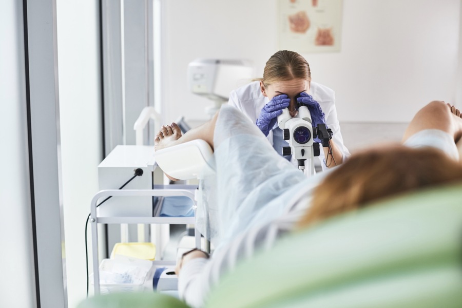
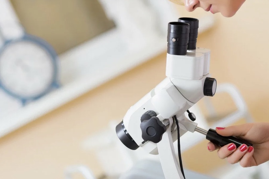
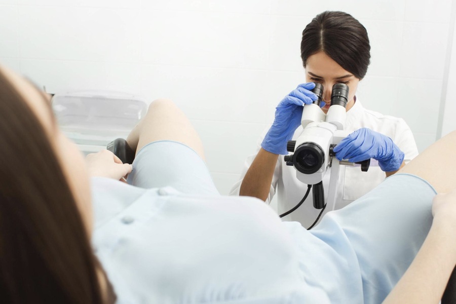
Colposcopy is a specialized diagnostic procedure designed to examine the cervix, vagina, and vulva in detail. Utilizing a colposcope, which is a binocular microscope with a light source, this method allows healthcare providers, including a top gynecologist in Dubai, to detect abnormalities in the mucous membranes and blood vessels. It is an essential tool in gynecology, contributing significantly to early diagnosis, prevention, and treatment of cervical cancer and other gynecological conditions.
Overview of Colposcopy
Purpose of the Procedure
The primary goal of colposcopy is to identify and evaluate changes in the cervical tissue that might indicate disease. By magnifying the cervical tissue and using diagnostic solutions, the procedure enhances the visibility of abnormalities that may not be apparent during a routine pelvic exam or Pap smear.
This examination is particularly useful for detecting precancerous and cancerous lesions, monitoring chronic conditions, and guiding biopsies when necessary.
The Role of the Colposcope
The colposcope is a non-invasive diagnostic instrument that magnifies the view of the cervix up to 40 times its normal size. It allows clinicians to observe surface irregularities, vascular patterns, and color changes that may indicate abnormal cellular activity.
Key Features
– Magnification: Adjustable settings for varying levels of detail.
– Lighting: High-intensity illumination for enhanced visibility.
– Non-contact: The device remains external, ensuring patient comfort.
Detailed Procedure
Preparation for Colposcopy
Before undergoing colposcopy, patients are typically advised to:
1. Schedule Appropriately
The procedure should be scheduled when the patient is not menstruating, as menstrual blood can obscure the examination.
2. Abstain from Vaginal Interference
Avoid intercourse, douching, and vaginal medications for 24-48 hours before the appointment to ensure optimal conditions for examination.
3. Relaxation Techniques
Anxiety about the procedure is common. Patients are encouraged to ask questions and practice relaxation techniques to ease tension.
Steps of the Procedure
1. Positioning
The patient lies on a gynecological chair with feet in stirrups, allowing access to the vaginal canal.
2. Insertion of the Speculum
A lubricated speculum is gently inserted to open the vaginal walls and provide a clear view of the cervix. This may cause mild pressure or discomfort.
3. Initial Observation
The clinician conducts a simple colposcopy, assessing the cervix’s size, shape, color, and vascular patterns.
4. Application of Diagnostic Solutions (for Extended Colposcopy)
Acetic Acid (3%): Applied to highlight abnormal areas. Abnormal cells temporarily appear white (acetowhite areas) under this solution.
Lugol’s Iodine: A solution containing iodine is used to identify glycogen-containing cells. Healthy squamous epithelium stains brown, while abnormal or glycogen-deficient cells remain unstained.
5. Magnified Examination
The colposcope is positioned at a distance of 10-15 cm from the genitalia, allowing a magnified view of the cervix without direct contact.
6. Biopsy (If Needed)
If suspicious areas are identified, a small tissue sample may be taken for further laboratory analysis. This process is quick but may cause slight cramping.
Duration and Sensation
The procedure typically takes 15-20 minutes. Most patients experience minimal discomfort:
Insertion of Speculum: May cause mild pressure.
Application of Solutions: Acetic acid or iodine might cause a brief burning or tingling sensation.

Types of Colposcopy
1. Simple Colposcopy
This involves a direct visual examination of the cervix without the use of diagnostic solutions. It provides an initial assessment of:
Cervical size and shape.
Mucosal surface and vascular patterns.
The transformation zone (where the squamous and columnar epithelium meet).
2. Extended Colposcopy
This includes the use of acetic acid and iodine to enhance the detection of abnormalities. Tissue reactions to these solutions help identify specific conditions and guide further treatment or biopsy.
Indications for Colposcopy
Colposcopy is recommended in various situations:
Preventive Screening
Women over 30 years of age are advised to undergo colposcopy annually as part of routine preventive care.
Factors increasing the need for regular screenings include:
Family history of cervical cancer.
Recurrent miscarriages.
Persistent HPV infection (especially high-risk types).
Long-term use of hormonal contraceptives without medical supervision.
Diagnostic Evaluation
Colposcopy is performed when abnormalities are detected during routine gynecological examinations, including:
Abnormal Pap smear results.
Positive HPV test (high-risk strains).
Symptoms such as:
Vaginal bleeding outside menstruation.
Chronic pelvic pain.
Unusual discharge.
Monitoring Known Conditions
Patients with diagnosed gynecological disorders, such as cervical dysplasia or HPV-related lesions, may require frequent colposcopies to:
Monitor disease progression.
Evaluate the effectiveness of treatments.
Pre- and Post-Operative Assessment
Colposcopy is essential in surgical planning and follow-up to ensure:
Accurate diagnosis before procedures.
Effective healing after interventions.
Conditions Diagnosed via Colposcopy
Colposcopy is instrumental in detecting and diagnosing a wide range of conditions, including:
1. Cervical Polyps
Benign growths that may cause bleeding or interfere with conception.
2. Condylomas
Wart-like lesions caused by HPV, often affecting the genital and anal regions.
3. Erosion and Pseudo-Erosion
Erosions caused by physical damage or irritation can progress to pseudo-erosions if untreated.
4. Endometriosis
Visible as purplish lesions caused by endometrial cells outside the uterus.
5. Dysplasia
Precancerous changes in the cervical cells, often appearing as white spots after acetic acid application.
6. Leukoplakia
White, keratinized patches associated with precancerous changes.
7. Cervical Scarring
Scars from childbirth or surgical procedures can deform the cervix, affecting menstruation and fertility.
8. Early-Stage Cancer
Colposcopy can detect subtle signs of cancer before they become invasive.

Advantages of Colposcopy
1. Early Detection of Cancer
Identifies precancerous and cancerous changes at an early stage, significantly improving treatment outcomes.
2. Guided Biopsy
Enables precise sampling of abnormal tissue for histological analysis.
3. Non-Invasive and Painless
The procedure involves minimal discomfort and is non-invasive unless a biopsy is required.
4. Quick and Efficient
The examination takes a short time and provides immediate insights.
Preparation and Aftercare
Before the Procedure
Schedule the procedure away from menstruation.
Avoid vaginal douching or intercourse 24-48 hours prior.
Wear comfortable clothing.
After the Procedure
Normal activities can typically be resumed immediately if no biopsy is performed.
If a biopsy is taken, mild spotting or cramping may occur for a day or two.
Conclusion
Colposcopy is a cornerstone of modern gynecological care. It provides unparalleled detail in diagnosing and managing cervical, vaginal, and vulvar conditions. Whether used for routine screening, diagnosis, or monitoring, colposcopy is an invaluable tool for protecting women’s health. Regular screenings and timely follow-ups can help detect abnormalities early, offering the best chances for effective treatment and a positive prognosis.

Soccer lover, vegan, DJ, Saul Bass fan and fullstack designer. Working at the crossroads of art and sustainability to craft delightful brand experiences. Let’s design a world that’s thoughtful, considered and aesthetically pleasing.
